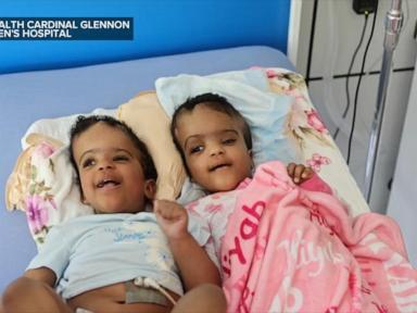
The successful separation of conjoined twins represents a convergence of advanced anatomical mapping, multidisciplinary surgical coordination, and rigorous postoperative physiological management. When conjoined twins undergo anatomical division, the clinical challenge extends far beyond the operating theater. It initiates a complex multi-system reorganization that tests the limits of pediatric critical care, vascular surgery, and metabolic stabilization. Understanding the true scope of such medical milestones requires dissecting the physiological dependencies, the surgical risk stratification, and the long-term rehabilitative framework necessary for independent survival.
The Anatomy of Shared Systems and Vascular Interdependence
Conjoined twins develop from a single fertilized ovum that fails to fully separate during the blastocyst stage, approximately 13 to 15 days post-fertilization. The clinical complexity of any separation procedure is dictated entirely by the degree of structural fusion and shared organ systems.
Craniopagus twins share cranial bones and cerebral vasculature; thoracopagus twins share the pericardium and myocardium; omphalopagus twins share the liver and gastrointestinal tract; ischiopagus twins share the pelvis, lower gastrointestinal tract, and urogenital systems. The primary determinant of surgical feasibility is not merely the presence of shared structures, but the functional independence and redundancy of those structures.
The vascular architecture frequently presents the highest intraoperative risk. Shared venous sinuses in craniopagus cases or fused hepatic parenchyma in thoracopagus and omphalopagus cases create high-flow vascular anastomoses. When surgeons divide these tissues, they manage acute hemodynamic shifts. The sudden closure or redistribution of blood volume alters preload, afterload, and cardiac output in both patients.
Hemodynamic Realignment and Cardiac Adaptation
Prior to separation, the cardiovascular systems of conjoined twins often operate as a single closed loop with bidirectional shunting. One twin may act as the physiological "donor," supplying blood and metabolic filtration to the other.
Upon division, each infant's cardiovascular system must independently maintain systemic perfusion, blood pressure, and tissue oxygenation.
- Preload Volatility: The sudden elimination of cross-circulation causes acute hypovolemia in the twin who was previously sustained by the shared vascular bed, while the other may experience fluid overload.
- Myocardial Strain: Ventricular function must rapidly adjust to the new systemic vascular resistance profile without the compensatory capacity of the sibling's circulatory volume.
- Autonomic Instability: Surgical manipulation near major neural pathways, particularly in craniopagus or thoraco-omphalopagus twins, frequently triggers transient autonomic dysregulation, manifested by severe bradycardia or tachyarrhythmias.
The Preoperative Decision Matrix and Imaging Modalities
Clinical teams deploy high-resolution imaging and three-dimensional modeling to construct an accurate roadmap before making a single surgical incision. Standard cross-sectional imaging is insufficient for navigating complex anatomical interfaces.
Magnetic resonance venography (MRV), computed tomography angiography (CTA), and high-resolution ultrasound provide precise visualization of vascular conduits.
- Virtual Surgical Planning: Teams utilize patient-specific three-dimensional printed replicas of shared organs to simulate the division plane and verify the preservation of critical blood supplies.
- Multidisciplinary Simulation: Surgical teams, anesthesiologists, perfusionists, and nursing staff conduct dry-run simulations to rehearse positioning, emergency hemorrhage control protocols, and the exact sequence of patient handoffs once physical separation occurs.
- Tissue Expansion: In cases involving extensive skin and soft tissue fusion, surgeons frequently deploy tissue expanders weeks or months prior to the primary procedure. This generates sufficient native skin coverage to achieve primary closure of the abdominal wall or cranium without tension, minimizing the risk of wound dehiscence or necrosis.
The Intraoperative Protocol and Staged Interventions
The separation procedure rarely occurs as a single, acute event. In complex presentations, teams employ a staged strategy to mitigate physiological shock and reduce cumulative anesthesia time.
Phase one typically involves the placement of tissue expanders and the gradual ligation of bridging blood vessels over multiple procedures. This encourages collateral vessel development and allows the microvasculature of each twin to adapt to altered flow dynamics prior to definitive division.
The definitive separation surgery requires parallel operating teams, often organized symmetrically within the same suite or in adjoining rooms prepared for simultaneous microvascular reconstruction.
- Anesthetic Management: Dual anesthesia teams manage independent endotracheal intubation, invasive arterial monitoring, central venous access, and continuous near-infrared spectroscopy (NIRS) to monitor cerebral and somatic tissue oxygenation.
- Anatomic Division Sequence: Hard tissues, such as fused pelvic bones or cranial plates, are divided using specialized osteotomes or ultrasonic bone scalpel technology to minimize thermal and mechanical trauma to underlying neural or vascular structures. Soft tissues are dissected using advanced energy devices that seal microvessels before division.
- Reconstructive Closure: Once physical separation is achieved, the secondary teams immediately assume care of each patient in parallel bays. Plastic and reconstructive surgeons execute complex flap rotations, biological mesh placement for abdominal wall reconstruction, or custom cranioplasty to protect exposed neural tissue.
Postoperative Critical Care and Metabolic Stabilization
The immediate postoperative window is characterized by intensive metabolic and respiratory management. The physiological stress of prolonged surgery induces a systemic inflammatory response syndrome (SIRS) that requires precise pharmacological and nutritional support.
Fluid management requires continuous monitoring of central venous pressure, lactate clearance, and hourly urine output. The newly independent bodies undergo profound catabolic shifts, necessitating early enteral or parenteral nutrition to prevent gut mucosal atrophy and support wound healing.
- Respiratory Mechanics: Infants with prior thoracic fusion frequently exhibit diaphragmatic dysfunction or chest wall instability, requiring prolonged mechanical ventilation and pulmonary hygiene protocols to prevent atelectasis and nosocomial pneumonia.
- Neuromuscular Rehabilitation: Prolonged immobilization during preoperative positioning and recovery demands structured physical and occupational therapy to address contractures, promote motor milestone attainment, and encourage independent neural pathway development.
- Psychological and Developmental Adaptation: Conjoined twins develop a unique sensorimotor and psychological bond characterized by constant physical proximity. Post-separation therapy addresses spatial orientation, body schema reorganization, and cognitive adaptation to independent physical existence.
Long-Term Prognosis and Longitudinal Tracking
Survival and functional outcomes depend directly on the specific anatomical system involved and the presence of associated congenital anomalies. While outcomes for omphalopagus and thoracopagus twins with separate hearts have improved significantly due to advances in neonatal critical care, craniopagus separations involving shared venous sinuses remain among the most challenging procedures in modern medicine, carrying substantial neurological morbidity risks.
Longitudinal care requires coordinated multidisciplinary oversight spanning pediatric subspecialties. Orthopedic surveillance tracks musculoskeletal alignment and spinal curvature; nephrological monitoring evaluates renal function following complex abdominal reconstruction; and neurodevelopmental assessments track cognitive progression.
The transition from a fused physiological state to functional independence marks the beginning of a multi-year rehabilitation trajectory. Success is measured not merely by the technical execution of the primary division, but by the sustained metabolic stability, structural integrity, and independent developmental progress of each patient over the course of childhood and into adulthood.